About:
Exploring new approaches to machine hosted
neural-network simulation, and the science
behind them.
Your moderator:
John Repici
A programmer who is obsessed with giving experimenters
a better environment for developing biologically-guided
neural network designs. Author of
an introductory book on the subject titled:
"Netlab Loligo: New Approaches to Neural Network
Simulation". BOOK REVIEWERS ARE NEEDED!
Can you help?
Other Blogs/Sites:
Neural Networks
Hardware (Robotics, etc.)
|
Monday, April 26. 2010
The improvements and innovations are coming fast and furious now. It is a great time to be into this stuff.
Traditional fMRI (it is strange to type that) detected blood flow. Hemoglobin in blood contains iron, which mediates changes in magnetic fields. These are, in turn, detected and converted to three-dimensional image data by the MRI. That same iron-containing molecule carries oxygen to the cells of the body. When neurons become more active they require more oxygen-carrying blood. Arteries in the vicinity of the active neuron cells respond by dilating in order to increase the supply. It is this extra blood that is detected by fMRI (traditional  fMRI, that is).
This has been a boon to understanding the topographical correlates of thought and brain function. That's the upside. The downside is that it produces very course correlates. That is, it only measures increased blood flow in the vicinity of neural activity. It doesn't pin-point the actual activity topographically.
It is also contextually course information. In other words, it displays all activity, and can't discern between, say long-, and short-term PTP, or any of the staggering number of other protein interactions that are involved in different types of mental activity. These different types of activity are often as important as locational activity.
Finally, it has a course temporal aperture as well. Because it measures blood-flow, it tends to see the activity many milliseconds, or even seconds after the activity has started
Some of the timing and delay deficiencies have been overcome by coupling it with EEG scans as well. EEG scans have almost no positional information, just giving general areas of the brain where electrical activity is sourced, but it does give immediate feedback, which can then be narrowed by the fMRI imaging that comes in some time later.
Now, the researchers at MIT have begun to work on new ways for the fMRI to image actual protein/neurotransmitter mediated activity in the brain. Instead of simply measuring the amount of activity through increased hemoglobin in the area, these will image on the actual molecules involved in brain functions. They are accomplishing this by coming up with contrast agents (things that make an MRI image brighter or darker), which bind directly to the various molecular sites and chemicals in the brain used in brain and neuron function.
The upshot? Faster, more tightly synchronized time windows, more fine-grained spacial resolutions and magnification scales, and a whole new dimension of functionality. The functionality is based on being able to contrast specific molecular mechanisms having to do with specific types of brain activity.
Tuesday, April 20. 2010
Proteins underlie, and enable most neural functionality. Our understanding of their three-dimensional structures, and how they operate continues to increase on an almost daily basis now. This study is one more step forward.
Our understanding of the role proteins play in memory and neural functioning is becoming increasingly more detailed as we gain understanding of how proteins, themselves, operate. This, in turn, is helping to clarify our ever more nuanced understanding of the memory mechanisms proteins support. In short, if you can read this, thank a protein molecule.
From the study:
"we found a modification to a protein that controls the cellular protein-synthesis machinery. This modification seems to affect the ability of nerve cells to communicate with each other and is thought to be part of the processes underlying memory."
Monday, April 5. 2010
In 1992 two men were awarded the Nobel Science Prize in Physiology or Medicine for work that is proving extremely important to our understanding of brain function at the cellular and molecular levels.
The 1992 prize went to Dr.s Edmond H. Fischer and Edwin G. Krebs for their research and discoveries of “reversible protein phosphorylation as a biological regulatory mechanism.” (i.e., glycogen phosphorylase).
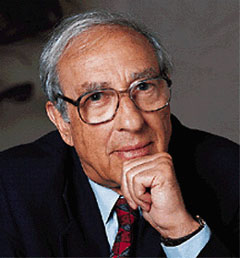
Dr. Edmond Fischer
|
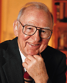
Dr. Edwin Krebs
|
Just a hunch, but I bet people get their names conflated every which way.
Monday, March 29. 2010
I'm not sure there's anything new here, other than demonstrating that something which has been confirmed by a variety of other means, does what you'd expect it to do in an fMRI machine. Pavlov referred to this phenomenon as the investigatory reflex.
This study shows that the reflex is represented in brain-activity as well as outward behavior. This gets us a little closer to the origin point, taking us a little farther down the path. In essence it shows us one more "observable" appearance of the investigatory reflex, which Pavlov had documented in the early nineteen-hundreds. That further validates and clarifies our understanding.
Notwithstanding the gee-whiz appeal of the fMRI, the real search—for me at least—is for the underlying mechanisms and brain characteristics that are responsible for the investigatory phenomenon. This particular study seems to have been restricted to only the appearance of the investigatory reflex in V1 (BA17).
This doesn't detract from the study's value, however. It isn't really fair for me, or anyone, to expect a custom fit to our questions in an "off-the-rack" world. This study should prove important, because it may help to show that the investigatory reflex has brain-global origins. That is, it may show that the investigatory reflex is a natural byproduct of individual neurons acting together, regardless of what brain-region they occupy. If not caused directly by local, cell-centric activities, the investigatory reflex may, at least, be facilitated by intrinsic cell behavior in some way. We are learning, for example, that the hippocampus seems to be tied into this reflexive phenomenon in a fairly substantial way.
I have some questions for the tailor: If the novel stimulus had been auditory, that probably would not have lit up V1 at all, but this is just a hunch on my part. It would have been interesting to find out—one way or the other—if V1 would have been lit up with novel auditory stimulus. This could lead to even more interesting questions about cross-modal stimuli. For example, what would happen at V1 if the novel auditory was sourced from specific locations around the organism? One interesting experiment along these lines might be to repeatedly play a given sound always from the same "location", and then suddenly (the novel part), make it emit from a different location.
Such questions simply aren't addressed in this study, which restricts itself, not only to just area V1, but also to just the effects of novel visual stimuli at V1. Also, there is no attempt made in the study to isolate the investigatory reflex at V1 from other areas that may have facilitated it. That is, the study doesn't really attempt to drill down and expose the brain structures and mechanisms actually responsible for the reflex. It is primarily about gathering more data-points, and there is nothing wrong with that.
- The Press Release: "The human brain processes predictable sensory input in a particularly efficient manner"
- From the more aptly named study: "Stimulus predictability reduces responses in primary visual cortex. - (abstract) - Full Text($)
- Background Knowledge:
Monday, March 22. 2010
A recent University of Minnesota study helps to clarify the role of the hippocampus in the formation and ongoing operations of long term memory. Specifically, with regard to the replay role that it has been know to participate in.
The hippocampus has long been known to aid in the playback of recent memories and has been thought to be a mechanism whereby recent, short-term memories could be repeated to aid in their transfer into long-term memory mechanisms within the brain.
The study demonstrated that, when faced with a novel challenge, the animals don't merely play back their most recent memories in their hippocampus activity patterns. Instead, it was found that they are most likely to play back experiences they had encountered least. Dr. Redish and his team also discovered that the animals often played back sequences that were never before experienced.
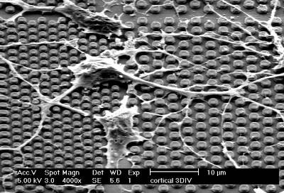
Tuesday, March 16. 2010
|
|
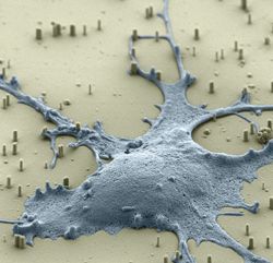
You wouldn't expect a good story on neural networks to come out of the nanotechnology crowd. Well, maybe you would. These two fields are fast converging. It wasn't that long ago that Carver Mead began constructing silicone based circuits that were designed to mimic exactly, what we knew about neurons in the retina. Now, one of his students, Kwabena Boahen, is working to use nanotechnology to more deeply investigate individual biological neurons. Is it just me, or does this look like somebody on the beach dropped their blue snow-cone.
|
|
|
Other Developments include
IMEC's micronail chips, upon which experimenters can grow biological cells, and test them.
|
Saturday, February 27. 2010
The title in the Journal of Experimental Biology is: BEES RECOGNISE FACES USING FEATURE CONFIGURATION. That's a bit of an over-reach, which was pushed even further out over the edge by the NYT reporting on the study.
I personally think there is a high probability that bees possess "facial recognition" facilities. This is just a hunch, but if bees have "facial recognition" facilities, wouldn't the "facial features" recognized look like bee faces, and not human faces? I'm just sayin'.
In any event, it is a good study because it does show rather convincingly that bees are able to recognize patterns composed of combinations of line-based features (the "faces" were stick-figure faces, with two dots for eyes, a slanted line for a nose, etc.).
|

|
 Stand Out Publishing


